Whether an Ultrasound Finds a Bichon Frise’s Stones
It's incredibly disheartening when your beloved Bichon Frise starts showing signs of urinary trouble—things like frequent squatting, straining to pee, or even accidents in the house. These worrying symptoms often point to bladder stones, a common and often recurrent issue for our fluffy friends. This guide will thoroughly explore whether an ultrasound is an effective tool for finding those pesky stones, what it involves, and how it fits into getting your Bichon Frise back to feeling their best.
The Role of Ultrasound in Diagnosing Bladder Stones
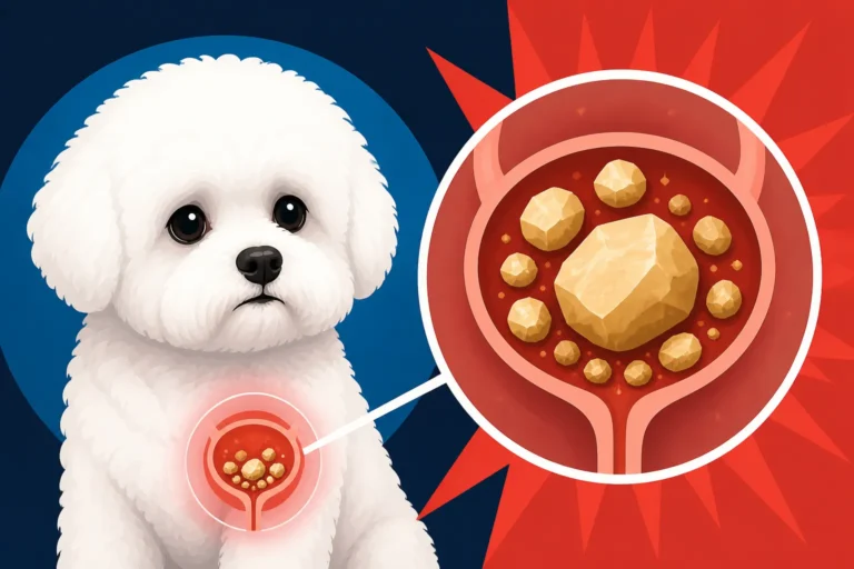
When your vet suspects bladder stones, the first images that usually come to mind are X-rays. And while X-rays are fantastic for seeing certain types of stones, they aren't the whole picture. This is where an ultrasound truly shines, offering a dynamic, real-time view that static X-rays simply can't provide. Many frustrated pet parents have experienced the confusion when X-rays come back clear, yet their dog's symptoms persist. This is often because not all stones are visible on an X-ray, leading to continued discomfort for your Bichon Frise.
Why Ultrasound Might Be Preferred Over X-rays Alone
You see, some types of bladder stones are "radiopaque," meaning they show up clearly on an X-ray because they block the X-ray beam. Think of calcium oxalate stones, which are unfortunately very common in Bichon Frises. These usually pop right out on a radiograph. However, other stones, like urate or cystine stones, are "radiolucent." They don't absorb X-rays well, if at all, and can be completely invisible on a standard X-ray film. This is a critical point, especially when your vet is trying to figure out what's really going on. An ultrasound uses sound waves, not radiation, and can visualize these radiolucent stones with remarkable clarity, essentially revealing the hidden culprits that X-rays might miss.
Imagine the relief you'd feel knowing that even if the first round of diagnostics didn't yield answers, there's another powerful tool available. This is particularly important because early detection can make a huge difference in treatment outcomes and preventing severe complications like a urethral blockage, which can be life-threatening. If your Bichon Frise is showing The First Signs a Bichon Frise Has Stones, an ultrasound could be a vital step in confirming the diagnosis.
What Ultrasound Reveals About Stones
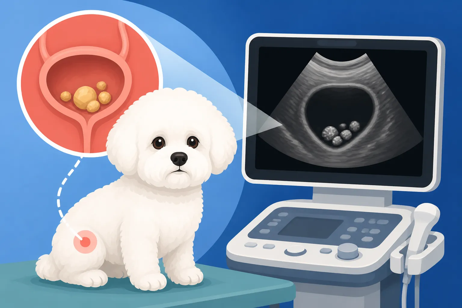
An ultrasound allows the veterinarian to see the internal structure of the bladder in detail. When stones are present, they typically appear as bright, reflective objects that cast an acoustic shadow. This shadow is a tell-tale sign of a solid object. The vet can often see multiple stones, their size, shape, and even if they are free-floating or adhered to the bladder wall. More than just seeing the stones, the ultrasound can also reveal associated issues, like inflammation of the bladder wall (cystitis) or the presence of thick sediment, which can sometimes be a precursor to stone formation.
It's a non-invasive procedure, meaning your Bichon Frise won't need surgery or injections for the imaging itself. Usually, they'll just need to lie calmly on their back, often in a comfortable "V-trough," while a small area of their belly is shaved and a gel applied. The vet then moves a transducer (a small handheld device) over the area, and images appear on a screen in real-time. This real-time aspect is fantastic because the vet can even watch the stones shift and move within the urine, confirming their presence and distinguishing them from other artifacts.
The Procedure: What to Expect During a Bichon Frise Bladder Ultrasound
Getting an ultrasound for your Bichon Frise is a fairly straightforward process, and understanding what happens can help ease your mind. It's often performed right in your vet's office or at a referral specialty clinic, depending on the equipment and expertise available. Unlike some procedures, it usually doesn't require general anesthesia, though mild sedation might be used for particularly anxious dogs to ensure they lie still.
Preparation for the Ultrasound
For the best possible images, there are a few preparatory steps. Typically, your vet will ask you to fast your Bichon Frise for about 8-12 hours before the appointment. This helps reduce gas in the intestines, which can interfere with the sound waves and obscure the view of the bladder and other abdominal organs. More importantly, they'll usually want your dog to have a moderately full bladder. This distends the bladder, making it easier to visualize any stones or abnormalities within its walls. Sometimes, they might even ask you to prevent your dog from urinating for a couple of hours before the appointment.
Once at the clinic, a small patch of fur on your Bichon Frise's lower abdomen will be gently shaved. This allows for direct contact between the ultrasound transducer and the skin, ensuring clear image transmission. A special ultrasound gel is then applied, which helps the transducer glide smoothly and eliminates air pockets that could interfere with the sound waves. Your Bichon Frise will then be positioned comfortably, usually on their back, and the diagnostic imaging begins. Many pet parents find the experience quite calm, especially if their dog is relaxed.
Interpreting the Ultrasound Images
During the ultrasound, the veterinarian or a veterinary radiologist will methodically scan the bladder and surrounding structures. They're looking for several key indicators:
- Stones and Sediment: As mentioned, stones appear as bright, hyperechoic structures with distinct acoustic shadowing. They'll also note any "sludge" or sediment, which is often a collection of crystals and cellular debris that can contribute to stone formation. This sediment can sometimes be mistaken for small stones or vice-versa, so an experienced eye is crucial.
- Bladder Wall Thickness: Chronic inflammation due to stones or infection can cause the bladder wall to thicken. The ultrasound provides precise measurements of this thickness, which can indicate the severity and duration of the problem.
- Lesions or Masses: While checking for stones, the vet will also be on the lookout for any other abnormalities, such as polyps or tumors growing on the bladder wall. These are far less common than stones but are important to identify.
- Kidney and Ureteral Health: Sometimes, bladder stones are just one piece of a larger puzzle. The vet will often extend the ultrasound to examine the kidneys and ureters (the tubes connecting the kidneys to the bladder). This helps determine if there's any 'backup' of urine (hydronephrosis) due to a blockage in the ureters or if stones have formed in the kidneys themselves. This holistic view is essential for a complete diagnosis and treatment plan.
This comprehensive imaging helps the vet answer not just "are there stones?" but also "how severe is the issue?" and "are other organs affected?" It's a critical tool in guiding the next steps, whether that's dietary management, medication, or surgery. For instance, understanding What a Calcium Oxalate Stone Is in a Bichon Frise is important, but locating it precisely with ultrasound is vital for successful removal.
Combining Ultrasound with Other Diagnostic Tools
While ultrasound is incredibly powerful, it's rarely used in isolation. Think of it as a crucial piece of a larger diagnostic puzzle. A thorough veterinary work-up for suspected bladder stones in a Bichon Frise will typically involve several tests, each providing unique information. This integrated approach ensures the most accurate diagnosis and the best possible treatment plan.
The Power of X-rays and Urinalysis Alongside Ultrasound
Often, a vet will start with a good old-fashioned X-ray, especially if they suspect radiopaque stones, which are very common in Bichons. X-rays give a great overview of the entire urinary tract, showing the number and general location of calcified stones. However, as we discussed, if those X-rays come back clear but your Bichon Frise is still straining, an ultrasound becomes indispensable for detecting those invisible, radiolucent stones. Think of it as a second pair of eyes, seeing what the first might have missed.
Alongside imaging, a urinalysis is paramount. This simple test provides a wealth of information about the urine itself: its pH (acidity or alkalinity), the presence of blood, protein, white blood cells, and most importantly, crystals. Different crystal types are associated with different stone compositions, offering vital clues even before stones are confirmed. For example, knowing Whether Blood in a Bichon Frise's Urine Means Stones from a urinalysis, coupled with an ultrasound, paints a much clearer picture. Blood tests, too, can be helpful in evaluating overall kidney function and metabolic health.
| Diagnostic Tool | What it Primarily Detects | Pros | Cons |
|---|---|---|---|
| X-ray | Radiopaque stones (e.g., calcium oxalate), stone count, overall bladder size/shape. | Quick, relatively inexpensive, good for initial screening of calcified stones. | Cannot see radiolucent stones (urate, cystine), static image, doesn't assess organ function or bladder wall. |
| Ultrasound | Radiolucent and radiopaque stones, sediment, bladder wall thickness, masses, kidney/ureteral health, real-time assessment. | Detects all stone types, provides detailed anatomical view, non-invasive, assesses surrounding tissues. | Operator dependent, can be more expensive than X-ray, gas can obscure views, does not show stone composition. |
| Urinalysis | pH, crystals, blood, protein, infection. | Quick, inexpensive, provides clues about stone type and infection. | Does not confirm stones or their location, can be misleading if sample is old or contaminated. |
This table clearly illustrates why a combination approach is so powerful. Each tool has its strengths and weaknesses, and together they provide a robust diagnostic framework for your Bichon Frise.
The Importance of Follow-Up and Stone Analysis
Finding the stones is a critical first step, but it’s not the end of the journey. Once stones are removed, either through surgery or via non-surgical methods like voiding hydropulsion, it's absolutely vital to have them analyzed. Stone analysis, performed by a specialized lab, determines the exact chemical composition of the stones. This information is a game-changer because it dictates the long-term management strategy, particularly the specific prescription diet your Bichon Frise will need to prevent recurrence.
Many Bichon Frises, unfortunately, are prone to forming stones repeatedly. It's a breed predisposition, and they can even form The Stone That Comes Back a Year After Bichon Frise Surgery. This is why analysis is so crucial. Without knowing the stone type, preventing future stones is like shooting in the dark. For instance, calcium oxalate stones require a very different preventive diet than urate stones. Therefore, even after a successful ultrasound and removal, the full diagnostic picture isn't complete until those stones have been analyzed. Pet owners often find that pet insurance policies, such as those from Healthy Paws, can be incredibly helpful in managing the costs associated with these follow-up diagnostics and the often lifelong management required.
Addressing Specific Concerns for Bichon Frise Owners
Bichon Frises have unique health considerations, and bladder stones are high on that list. Their predisposition to certain types of stones, their small size, and the potential for recurrence mean that vigilant care and appropriate diagnostic tools are essential. Understanding these breed-specific nuances can help you advocate effectively for your pet's health.
Why Bichon Frises are Prone to Stones and Ultrasound's Specific Value
It's a frustrating reality that Bichon Frises are statistically more prone to developing bladder stones, particularly calcium oxalate stones. In fact, they are among the breeds that top the stone risk list. This inherited predisposition means that any signs of urinary discomfort should be taken very seriously. An ultrasound's ability to clearly visualize even small stones, identify sediment, and assess the overall health of the bladder lining is especially valuable for this breed. Early detection through ultrasound can mean the difference between managing a few small stones with a The Diet a Bichon Frise Stays On for Life and facing an emergency surgical procedure for a severe blockage.
Imagine your little Bichon constantly needing to go outside, straining, or having accidents. These are clear indicators that something is amiss, and waiting could escalate the problem. For a breed so prone to these issues, having a diagnostic tool like ultrasound that provides such a detailed, non-invasive look is a tremendous advantage. It allows vets to pinpoint problems that might otherwise remain hidden, giving you and your pet the best chance at a successful outcome. Pet parents often rely on pet insurance like Healthy Paws to cover advanced diagnostics like ultrasounds when these breed-specific issues arise, helping to alleviate the financial stress during an already worrying time.
Monitoring and Long-Term Management
For Bichon Frises with a history of bladder stones, ongoing monitoring is absolutely key. Ultrasound plays a significant role here, too. After initial treatment, your vet may recommend periodic bladder ultrasounds, sometimes annually or even every six months, especially if your dog has experienced recurrent stones. This allows them to catch new stone formation while the stones are still small and easier to manage, often before they cause severe symptoms or lead to a dangerous blockage.
It's about being proactive rather than reactive. Instead of waiting for the tell-tale signs of discomfort to reappear, regular check-ups that include an ultrasound can help you stay ahead of the problem. This type of proactive approach can significantly improve your Bichon Frise's quality of life and potentially reduce the need for repeat surgeries. While Whether a Bichon Frise Should Be X Rayed Yearly is a common question, many vets will opt for ultrasound in rotation or as the primary imaging method for high-risk individuals, especially if their previous stones were radiolucent. Discussing these preventative measures and associated costs, which can often be mitigated by comprehensive pet insurance like Healthy Paws, is an important conversation to have with your veterinarian.
When to Seek Emergency Ultrasound
Sometimes, symptoms escalate rapidly. If your male Bichon Frise suddenly cannot urinate, is straining intensely, or showing signs of extreme pain or lethargy, it’s an emergency. This could indicate a complete urethral blockage, which is life-threatening. In such cases, an emergency ultrasound is often performed immediately to confirm the blockage, identify the location of the stone (or stones), and assess the extent of the damage to the bladder and kidneys. Time is critical when Whether a Blocked Bichon Frise Can Die is a real concern. The speed and detail offered by ultrasound can be invaluable in these urgent situations, helping the veterinary team determine the quickest and most effective course of action, which often involves emergency surgery.
Disclaimer
The information provided in this article is for general educational purposes only and is not a substitute for professional veterinary advice. Always consult with a qualified veterinarian for any health concerns or before making any decisions about your pet's health or treatment plan.